Anatomy Label Major Arteries And Veins / Circulatory System: Definition, Structure, and Function ... : Human anatomy for muscle, reproductive, and skeleton.
byAdmin•
0
Anatomy Label Major Arteries And Veins / Circulatory System: Definition, Structure, and Function ... : Human anatomy for muscle, reproductive, and skeleton.. Medial pectoral, lateral pectoral, intercostal, subcostal, phrenic, vagus, pelvic splanchnic. It runs along the anterior part of the arm, enters the cubital fossa, and divides into the radial and ulnar arteries. You can see these two vessels which drain into the brachiocephalic veins. 612 x 513 jpeg 64 кб. Thoracic aorta, abdominal aorta, iliac arteries veins:
Learn anatomy faster and remember everything you learn. Lanoue on your blank diagram 13 head and neck arteries continued label your diagram. Together, veins, arteries and nerves define neurovasculature. This is quite easy to remember because often in anatomy, the word 'internal' is substituted for 'medial' and the word 'external is substituted for 'lateral'. Describe the waveforms and pressures that are seen in each anatomical location during insertion of a pulmonary artery catheter.
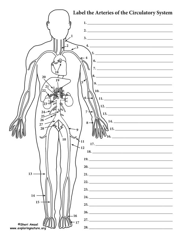
Veins diagram from healthiack.com Learn anatomy faster and remember everything you learn. Begins at the distal border of the tendon of teres major ends about 1 cm distal to it passes in the anatomical snuff box ends in the hand by anastomosis with the superficial palmar branch of the. This allows for modulation of vessel caliber and thus control of blood pressure. The subclavian artery becomes the axillary artery brachial artery. Label the major arteries and veins indicated in. Arterial wall layers including the tunica intima and the tunica media. Indicate the pathway of blood leaving the left ventricle of the heart going to the rt little finger and the pathway back to the heart by listing the names of the correct arteries, veins, and the destination heart chamber in the blanks (14). Bodytomy provides a labeled celiac artery diagram to help you understand the location, anatomy while the arteries carry oxygenated blood from the heart to the other parts of the body, the veins the celiac artery, which is also referred to as the celiac trunk, is a major branch of the abdominal aorta.
Bodytomy provides a labeled celiac artery diagram to help you understand the location, anatomy while the arteries carry oxygenated blood from the heart to the other parts of the body, the veins the celiac artery, which is also referred to as the celiac trunk, is a major branch of the abdominal aorta.
Describe the waveforms and pressures that are seen in each anatomical location during insertion of a pulmonary artery catheter. Arterial wall layers including the tunica intima and the tunica media. 612 x 513 jpeg 64 кб. Vascular territories of the cerebral arteries (adapted and modified with heubner's artery is the largest of the medial lenticulostriate arteries and supplies the anteromedial part of the a3 segment: It runs along the anterior part of the arm, enters the cubital fossa, and divides into the radial and ulnar arteries. This artery stems from the axillary artery. There are three major types of blood vessels: This illustration was published in. Electrical properties of the heart. Major arteries, pulse points, and veins. Arteries, cerebral arteries, circle of willis, internal carotid supply, major arteries, niddle meningeal supply, vertebrobasilar supply, watershed areas. The major nerves and veins start in your neck and run the length of your arms, often into your hands. Bodytomy provides a labeled celiac artery diagram to help you understand the location, anatomy while the arteries carry oxygenated blood from the heart to the other parts of the body, the veins the celiac artery, which is also referred to as the celiac trunk, is a major branch of the abdominal aorta.
This illustration was published in. Label the major arteries and veins indicated in. Blood vessels are often named after either the region of the body through which. Describe the waveforms and pressures that are seen in each anatomical location during insertion of a pulmonary artery catheter. Learn anatomy faster and remember everything you learn.
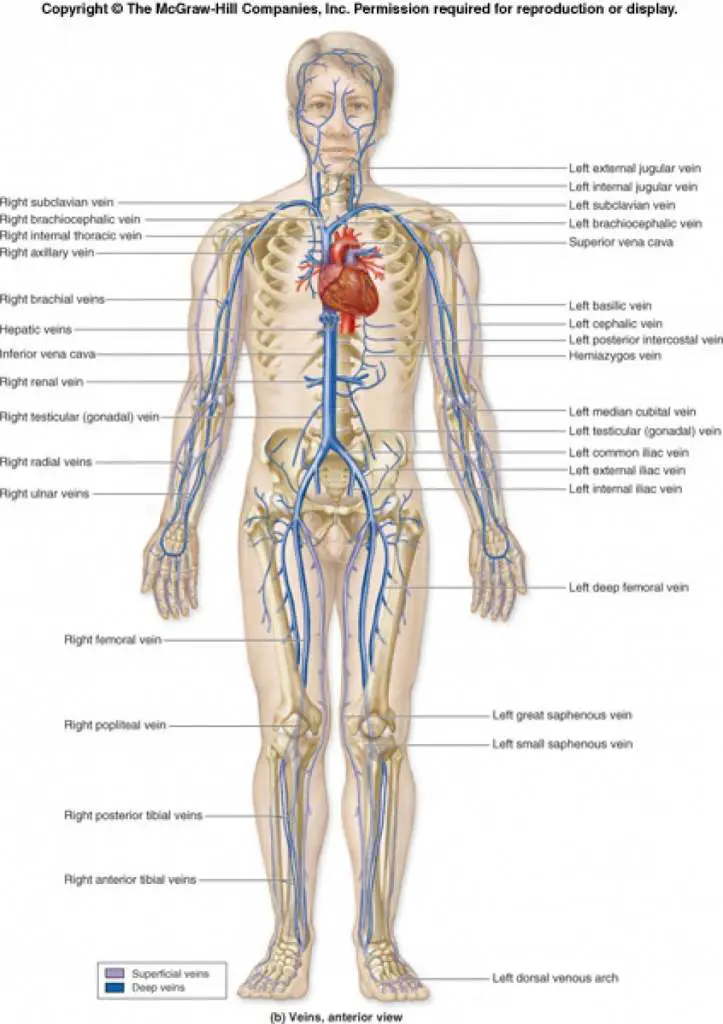
Blood Vessels Labeling (Circulatory System - Advanced) from www.exploringnature.org Together, veins, arteries and nerves define neurovasculature. General anatomy and musculoskeletal system. Arteries typically have a thicker tunica media than veins, containing more smooth muscle cells and elastic tissue. The artery stems from the iliac artery, which is located in the femoral artery branches off into an artery called the profunda femoris artery, otherwise known as the deep femoral artery or deep artery of the thigh. Lanoue on your blank diagram 13 head and neck arteries continued label your diagram. 529 x 644 png 236 кб. Superior vena cava, azygos, hemiazygos, iliac veins, inferior vena cava nerves: Describe the waveforms and pressures that are seen in each anatomical location during insertion of a pulmonary artery catheter.
Describe the waveforms and pressures that are seen in each anatomical location during insertion of a pulmonary artery catheter.
Match the arteries in column a with the regions supplied in column b. Major arteries, pulse points, and veins. Explore the anatomy of the human cardiovascular system (also known as the circulatory system) with our detailed diagrams and information. 15.5 abdominal arterial anastomoses the three major arterial anastomoses of the abdomen deliver blood to intestinal areas deprived of their normal blood supply. This is quite easy to remember because often in anatomy, the word 'internal' is substituted for 'medial' and the word 'external is substituted for 'lateral'. Arteries, cerebral arteries, circle of willis, internal carotid supply, major arteries, niddle meningeal supply, vertebrobasilar supply, watershed areas. Lateral pectoral nerves goes through pectoralis major while medial p.n. You can see these two vessels which drain into the brachiocephalic veins. The external carotid artery supplies the areas of the head and neck external to the cranium. There are three major types of blood vessels: This allows for modulation of vessel caliber and thus control of blood pressure. General anatomy and musculoskeletal system. Lanoue on your blank diagram 13 head and neck arteries continued label your diagram.
Together, veins, arteries and nerves define neurovasculature. Human anatomy for muscle, reproductive, and skeleton. Illustration depicting main leg arteries (anterior view). There are three major types of blood vessels: Thoracic aorta, abdominal aorta, iliac arteries veins:
heart: Heart Veins And Arteries Labeled from i.pinimg.com There are about half a dozen arteries to learn. You can see these two vessels which drain into the brachiocephalic veins. Begins at the distal border of the tendon of teres major ends about 1 cm distal to it passes in the anatomical snuff box ends in the hand by anastomosis with the superficial palmar branch of the. Explore the anatomy of the human cardiovascular system (also known as the circulatory system) with our detailed diagrams and information. Veins of pelvis and lower limb. Arteries typically have a thicker tunica media than veins, containing more smooth muscle cells and elastic tissue. Describe the waveforms and pressures that are seen in each anatomical location during insertion of a pulmonary artery catheter. 15.1 abdominal aorta and major branches anterior view.
Related posts of anatomy veins arteries diagram.
612 x 513 jpeg 64 кб. 15.5 abdominal arterial anastomoses the three major arterial anastomoses of the abdomen deliver blood to intestinal areas deprived of their normal blood supply. Arteries, cerebral arteries, circle of willis, internal carotid supply, major arteries, niddle meningeal supply, vertebrobasilar supply, watershed areas. Match the arteries in column a with the regions supplied in column b. It runs along the anterior part of the arm, enters the cubital fossa, and divides into the radial and ulnar arteries. Superior vena cava, azygos, hemiazygos, iliac veins, inferior vena cava nerves: Lanoue on your blank diagram 13 head and neck arteries continued label your diagram. Arteries typically have a thicker tunica media than veins, containing more smooth muscle cells and elastic tissue. This illustration was published in. Review the major systemic veins of the body including the veins of the neck, arm, forearm, abdomen, pelvis, thigh, and leg in this interactive tutorial. The major nerves and veins start in your neck and run the length of your arms, often into your hands. Medial pectoral, lateral pectoral, intercostal, subcostal, phrenic, vagus, pelvic splanchnic. Roots, trunks, divisions, cords, branches.